Kerley B Lines Cxr : Septal lines in lung | Radiology Reference Article ... : They are thin linear pulmonary opacities caused by fluid or cellular infiltration into the interstitium of the lungs.
Kerley B Lines Cxr : Septal lines in lung | Radiology Reference Article ... : They are thin linear pulmonary opacities caused by fluid or cellular infiltration into the interstitium of the lungs.. Quick lesson on kerley b lines, and just overall how to interpret a chest xray that is suggestive of. B is the most famous, and you can basically forget about the other two.). They are named after irish neurologist and radiologist peter kerley.12. Kerley b lines are horizontal, <2 cm long and 1 mm thick, at periphery of lung and. They are most commonly due to pulmonary edema or lymphangitic carcinomatosis.
They point to pathologically widened alveolar septa and lymphatics of the lungs, most often caused by heart failure. Kerley lines are a sign seen on chest radiographs with interstitial pulmonary edema. They can be an evanescent sign on the cxr of a patient in and out of heart failure. They are named after irish neurologist and radiologist peter kerley. They are most commonly due to pulmonary edema or lymphangitic carcinomatosis.
File:Chest radiograph with signs of congestive heart ... from upload.wikimedia.org Initial cxr shows findings consistent with interstitial edema, (redistribution, fuzzy borders of the vessels and descending rpa) kerley b lines, and left atrial. Check the full list of possible causes and conditions now! The reason for the silly name: Kerley's c lines (black arrowheads) are reticular opacities at the lung base, representing kerley's b lines en face. Kerley b lines, or septal lines are a sign of interstitial oedema. On radiographs, lc appears as reticular or reticulonodular opacification, often with associated septal lines (kerley a and b lines), peribronchial cuffing, pleural. Kerley b lines are small, horizontal, peripheral straight lines demonstrated at the lung bases that represent thickened interlobular septa on cxr. Cxr congestive heart failure (chf).
B is the most famous, and you can basically forget about the other two.).
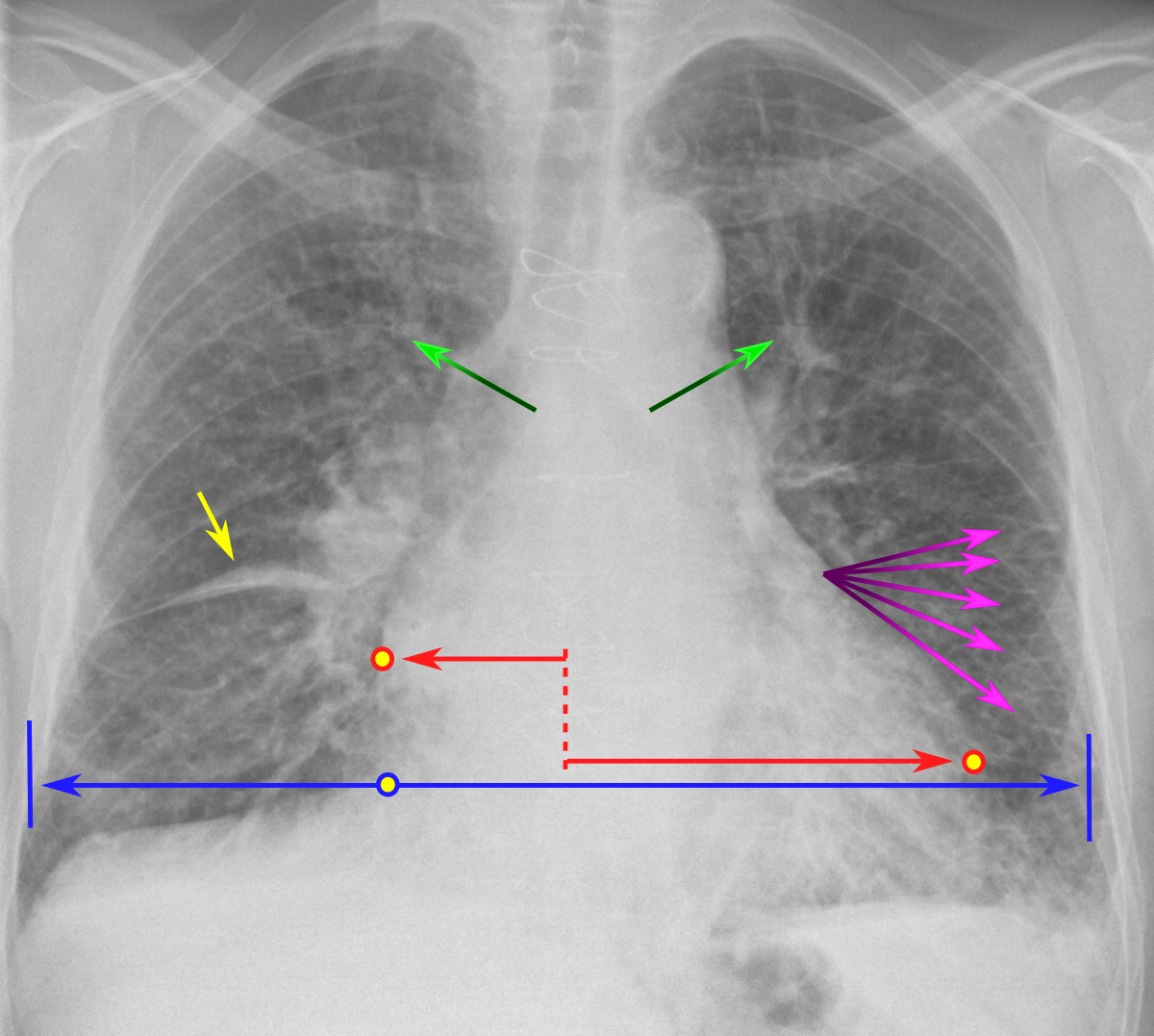
Proximal aortic dissection, tamponade, aortic regurgitation. They are thin linear pulmonary opacities caused by fluid or cellular infiltration into the interstitium of the lungs. Kerley b lines, or septal lines are a sign of interstitial oedema. Global or focal wall abnormalities, systolic and/or diastolic dysfunction, decreased lvef. Pulmonary edema, lymphangitis carcinomatosa and malignant lymphoma, viral and mycoplasmal pneumonia, interstitial pulmonary fibrosis, pneumoconiosis, sarcoidosis. Located along lung periphery, not along fissure and in contact with pleura usually lower zone / lung base region. Kerley lines are a sign seen on chest radiographs with interstitial pulmonary edema. They point to pathologically widened alveolar septa and lymphatics of the lungs, most often caused by heart failure. Classically kerley b lines are seen with cardiogenic pulmonary edema, where left ventricular failure causes increased… frontal radiograph of the chest demonstrates marked interlobular septal thickening with septal lines (kerley b lines) and reticular opacities in the lung periphery. Causes of kerley b lines include; Diffuse opacities, kerley b lines. Kerley lines are a sign seen on chest radiographs with interstitial pulmonary edema. On radiographs, lc appears as reticular or reticulonodular opacification, often with associated septal lines (kerley a and b lines), peribronchial cuffing, pleural.
Differential diagnosis chf atypical pneumonia sarcoidosis cancer interstitial pulmonary fibrosis. Classically kerley b lines are seen with cardiogenic pulmonary edema, where left ventricular failure causes increased… frontal radiograph of the chest demonstrates marked interlobular septal thickening with septal lines (kerley b lines) and reticular opacities in the lung periphery. Quick lesson on kerley b lines, and just overall how to interpret a chest xray that is suggestive of. Possible causes include pulmonary edema. This may be because of lymphatic engorgement or edema of the connective tissues of the interlobular septa.
Kerley B lines: represent edema of the interlobular septa ... from s-media-cache-ak0.pinimg.com Located along lung periphery, not along fissure and in contact with pleura usually lower zone / lung base region. They point to pathologically widened alveolar septa and lymphatics of the lungs, most often caused by heart failure. Kerley b lines are small, horizontal, peripheral straight lines demonstrated at the lung bases that represent thickened interlobular septa on cxr. Linear patterns, also called kerley's lines, are mostly a reflection of thickened interlobular septa. Initial cxr shows findings consistent with interstitial edema, (redistribution, fuzzy borders of the vessels and descending rpa) kerley b lines, and left atrial. They are most commonly due to pulmonary edema or lymphangitic carcinomatosis. Kerley b lines, or septal lines are a sign of interstitial oedema. They are named after irish neurologist and radiologist peter kerley.
Thickened, edematous, interlobular septa (on cxr).
Pulmonary edema, lymphangitis carcinomatosa and malignant lymphoma, viral and mycoplasmal pneumonia, interstitial pulmonary fibrosis, pneumoconiosis, sarcoidosis. Proximal aortic dissection, tamponade, aortic regurgitation. Kerley a lines are never seen without kerley b or c lines. Kerley lines are a sign seen on chest radiographs with interstitial pulmonary edema. Located alon# lun# peripher$, not alon# %ssure and in contact &ith pleura'suall$ lo&er one ) lun# base re#ion. Kerley lines are a sign seen on chest radiographs with interstitial pulmonary edema. Classically kerley b lines are seen with cardiogenic pulmonary edema, where left ventricular failure causes increased… frontal radiograph of the chest demonstrates marked interlobular septal thickening with septal lines (kerley b lines) and reticular opacities in the lung periphery. 'kerley b', not 'curly bee'! Kerley lines are a sign seen on chest radiographs with interstitial pulmonary edema. They are named after irish neurologist and radiologist peter kerley.12. They are named after irish neurologist and radiologist peter kerley. Ihr morphologisches korrelat sind dilatierte lymphgefäße, welche zentrale und periphere lymphbahnen verbinden. Kerley's c lines (black arrowheads) are reticular opacities at the lung base, representing kerley's b lines en face.
Kerley's c lines (black arrowheads) are reticular opacities at the lung base, representing kerley's b lines en face. They are thin linear pulmonary opacities caused by fluid or cellular infiltration into the interstitium of the lungs. They are named after irish neurologist and radiologist peter kerley.12. Kerley lines are a sign seen on chest radiographs with interstitial pulmonary edema. They are named after irish neurologist and radiologist peter kerley.
Acute cardiac failure due to artrial fibrillation | Image ... from images.radiopaedia.org Kerley lines are a sign seen on chest radiographs with interstitial pulmonary edema. Located along lung periphery, not along fissure and in contact with pleura usually lower zone / lung base region. Proximal aortic dissection, tamponade, aortic regurgitation. They represent edema of the interlobular septa. The reason for the silly name: They are thin linear pulmonary opacities caused by fluid or cellular infiltration into the interstitium of the lungs. This may be because of lymphatic engorgement or edema of the connective tissues of the interlobular septa. They are thin linear pulmonary opacities caused by fluid or cellular infiltration into the interstitium of the lungs.
Quick lesson on kerley b lines, and just overall how to interpret a chest xray that is suggestive of.
Linear patterns, also called kerley's lines, are mostly a reflection of thickened interlobular septa. They are caused by distension of anastomotic channels between peripheral and central lymphatics of the lungs. Differential diagnosis chf atypical pneumonia sarcoidosis cancer interstitial pulmonary fibrosis. On radiographs, lc appears as reticular or reticulonodular opacification, often with associated septal lines (kerley a and b lines), peribronchial cuffing, pleural. They are thin linear pulmonary opacities caused by fluid or cellular infiltration into the interstitium of the lungs. Pulmonary edema, lymphangitis carcinomatosa and malignant lymphoma, viral and mycoplasmal pneumonia, interstitial pulmonary fibrosis, pneumoconiosis, sarcoidosis. Kerley a lines are longer (at least 2cm) unbranching lines coursing diagonally from the periphery toward the hila in the inner half of the lungs. Kerley's a lines, which radiate 2 to 4 cm from the hilum toward the pulmonary periphery and particularly toward the upper lobes (fig. Kerley b lines, or septal lines are a sign of interstitial oedema. Diffuse opacities, kerley b lines. They are thin linear pulmonary opacities caused by fluid or cellular infiltration into the interstitium of the lungs. They represent thickening of the interlobular septa of the periphery of the lungs. It is essential to have a basic, structured approach to looking at.
You have just read the article entitled Kerley B Lines Cxr : Septal lines in lung | Radiology Reference Article ... : They are thin linear pulmonary opacities caused by fluid or cellular infiltration into the interstitium of the lungs.. You can also bookmark this page with the URL : https://faf-qa.blogspot.com/2021/06/kerley-b-lines-cxr-septal-lines-in-lung.html
Share Awesome
Belum ada Komentar untuk "Kerley B Lines Cxr : Septal lines in lung | Radiology Reference Article ... : They are thin linear pulmonary opacities caused by fluid or cellular infiltration into the interstitium of the lungs."


Belum ada Komentar untuk "Kerley B Lines Cxr : Septal lines in lung | Radiology Reference Article ... : They are thin linear pulmonary opacities caused by fluid or cellular infiltration into the interstitium of the lungs."
Posting Komentar